


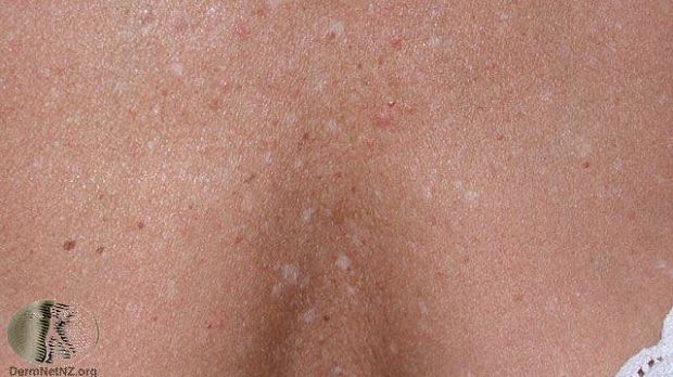
What Happened to the Melanocytes?
Idiopathic guttate hypomelanosis (IGH) is a pigmentary disorder characterized by small discrete white macules. The lesions have reduced amounts of melanin and few melanocytes. The pathogenesis of IGH remains unknown, but some studies have suggested the UV light can affect its progression. Melanin synthesis happens within melanocytes, and skin pigmentation is due to combination of functions of melanocyte and keratinocytes.
A recent study looked at the role of melanocytes in the pathogenesis of IGH. The study enrolled six patients and six control patients and cultured melanocytes in both groups. Then each culture was checked for melanin content. The results showed that melanin accumulated more in the basement layer of IGH patients, compared to the control. In addition, melanin distribution was uniform in control skin, while IGH patients showed patches of high and low melanin content. The melanocytes usually distribute melanin to keratinocytes, but in IGH patients this occurred in a non-uniform manner. Also, melanocytes from IGH patients were bigger in size and had more melanin than the control melanocytes.
The authors conclude that communication of the melanocytes to the dendrites that distribute pigment to the keratinocytes is impaired in the IGH patients. They state that they observed smaller and retracted dendrites in the IGH melanocytes in their patients. This may reduce the transfer of melanin from melanocytes to keratinocytes which may lead to the accumulation of more melanin and ultimately leads to deterioration in the IGH melanocytes.
Byline: Martha L. Sikes, MS, RPh, PA-C
Posted: May 21, 2018
Source: Wiley Online Library
Adapted from the original article.
[Image: DermNet New Zealand]